Functions of the Lymphatic System:
- Fluid Balance
- Fat absorption
- Defense
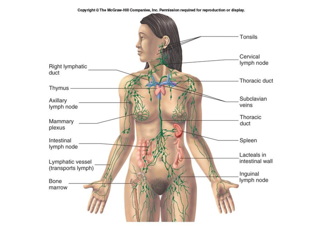
Overview of the Lymphatic System:
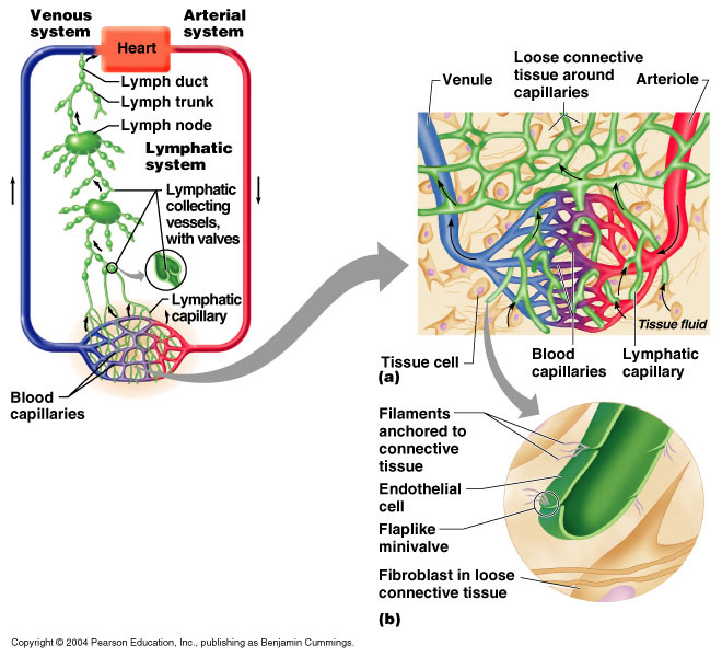
- Lymphatic capillaries remove fluid from tissues. The fluid becomes lymph.
- Lymph flows through lymphatic vessels, which have valves that prevent the back flow of lymph.
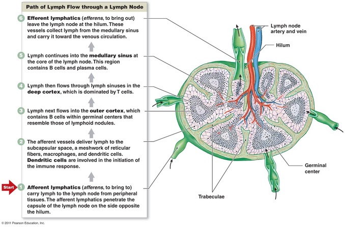
- Lymph nodes filter lymph and are sites where lymphocytes respond to infections.
- Lymph enters the thoracic duct or the right lymphatic duct.
- Lymph enters the blood.
- Lacteals in the small intestine absorb fats, which enter the thoracic duct.
- Chyle, which is lymph containing fats, enters the blood.
- The spleen filters blood and is a site where lymphocytes respond to infections.
- Lymphocytes (pre-B and pre-T cells) originate from stem cells in the red bone marrow. The pre-B cells become mature B cells in the red bone marrow and are released into the blood. The pre-T cells enter the blood and migrate to the thymus.
- The thymus is where pre-T cells derived from red bone marrow increase in number and become mature T cells that are released into the blood.
- B cells and T cells from the blood enter and populate all lymphatic tissues. These lymphocytes can remain in tissues or pass through them and return to the blood. B cells and T cells can also respond to infections by dividing and increasing in number. Some of the newly formed cells enter the blood and circulate to other tissues.
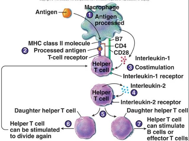
Proliferation of Helper T Cells:
- Antigen-presenting cells, such as macrophages, phagocytize, process, and display antigens on the cell's surface.
- The antigens are bound to MHC class II molecules, which present the processed antigen to the T-cell receptor of the helper T cell.
- Costimulation results from interleukin-1, secrete by the macrophage, and the CD4 glycoprotein of the helper T cell.
- Interleukin-1 stimulates the helper T cell to secrete interleukin-2 and to produce interleukin-2 receptors.
- The helper T cell stimulates itself to divide when interleukin-2 binds to interleukin-2 receptors.
- The "daughter" helper T cells resulting from this division can be stimulated to divide again if they are exposed to the same antigen that stimulated the "parent" helper T cell. This greatly increases the number of helper T cells.
- The increased number of helper T cells can facilitate the activation of B cells or effector T cells.
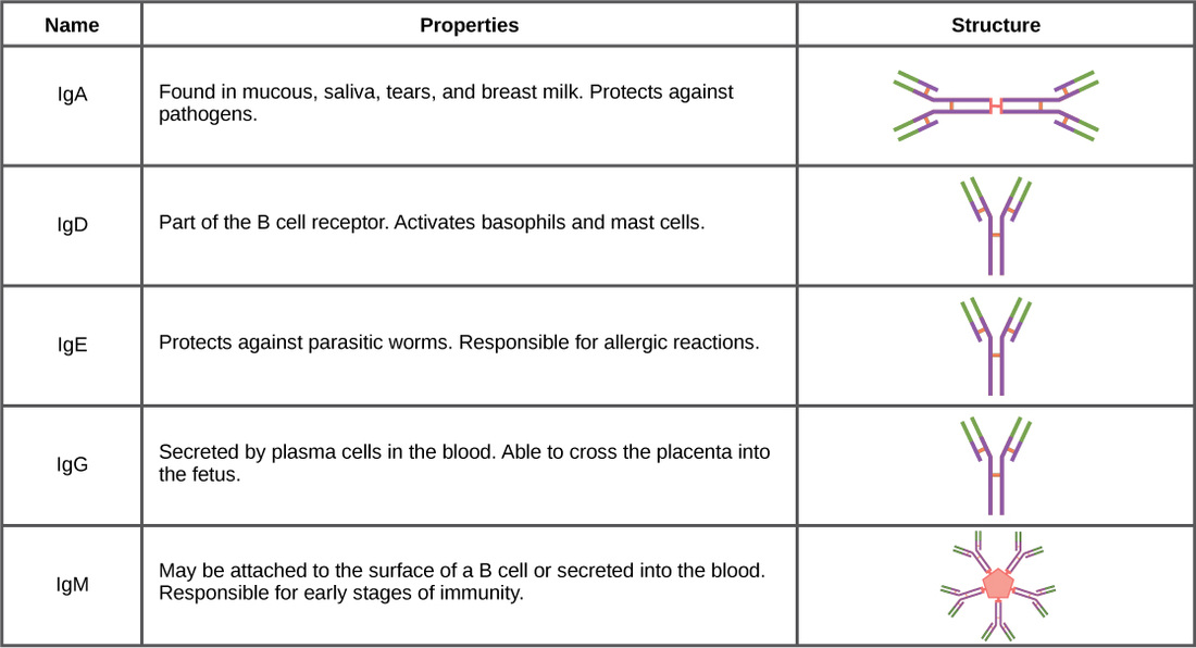
Classes of Antibodies and their functions
Proliferation of Cytotoxic T Cells:
- An MHC class I molecule displays an antigen, such as viral protein, on the surface of a target cell.
- The activation of cytotoxic T cell begins when the T-cell receptor binds to the MHC class I/antigen complex.
- There is costimulation of the cytotoxic T cell by CD8 and other surface molecules.
- There is costimulation by cytokines, such as interleukin-2, released from helper T cells.
- The activated cytotoxic T cell divides, the resulting daughter cell divide, and so on, eventually pro ducting many cytotoxic T cells.
 RSS Feed