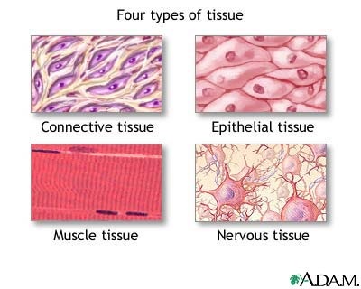
A tissue is a group of cells with similar structure and function that have similar extracellular substances located between them. The microscopic study of tissue structure is called histology. The four basic tissue types are epithelial, connective, muscle, and nervous.
Epithelial Tissue. Epithelial tissue (covering or lining), covers external and internal surfaces throughout the body. Surfaces of the body include the outer layer of the skin and the lining of cavities, such as the digestive tract, respiratory passages, and blood vessels. It also forms most glands. Epithelium consists almost entirely of cells with very little extracellular space between them. Although there are some exceptions, most epithelia have a free surface, which is not in contact with other cells, and a basal surface adjacent to a basement membrane, which attaches the epithelial cells to underlying tissues.
Functions of the epithelia are: protecting underlying structures, acting as a barrier, permitting the passage of substances, secreting substances, and absorbing substances.
Functions of the epithelia are: protecting underlying structures, acting as a barrier, permitting the passage of substances, secreting substances, and absorbing substances.
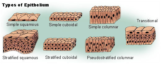
Epithelia are classified according to the number of cell laters and the shape of the cells.
Number of Layers Cell Shape
Simple (one layer) Squamous
Cuboidal
Columnar
Pseudostratified (a modified form of simple epithelium) Columnar
-------------------------------------------------------------
Stratified (more than one layer) Squamous
Cuboidal
Columnar
Transitional (a type of stratified epithelium)
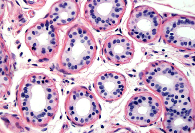
Simple Cuboidal Epithelium.
Function: Active transport and facilitated diffusion result in secretion and absorption by cells of the kidney tubules; secretion by cells of glands and choroid plexuses; movement of particles embedded in mucus out of the terminal bronchioles by ciliated cells.
Location: Kidney tubules, glands and their ducts, choroid plexuses of the brain, lining of terminal bronchioles of the lungs, and surfaces of the ovaries.
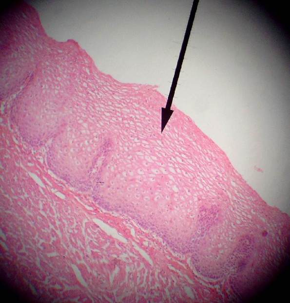
Stratified Squamous Epithelium (non-keratinized)
Function: Protects against abrasion, forms a barrier against infection, and reduces loss of water from the body.
Location: Keratinized - outer later of the skin; Non-Keratinized- mouth, throat, larynx, esophagus, anus, vagina, inferior urethra, and corneas.
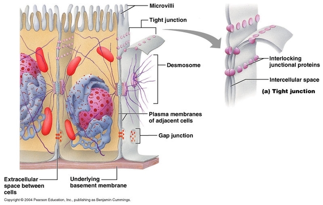 Cell Connections
 Epithelial cells are connected to one another in several ways.
Tight Junctions- bind adjacent cells together and form permeability barriers. Tight junctions prevent the passage of materials between epithelial cells because they completely surround each cell, similar to the way a belt surrounds the waist. Materials that pass throughout he epithelial later must pass through the cells, so those cells regulate what materials can cross. Tight junctions are found in the lining of the intestines and in most other simple epithelia.
Desmosomes- mechanical links that bind cells together. Modified desmosomes, called hemidesmosomes also anchor cells to the basement membrane. Many desmosomes are found in epithelia subjected to stress, such as the stratified squamous epithelium of the skin.
Gap Junctions- Small channels that allow small molecules and ions to pass from one epithelial cell to an adjacent one. Most epithelial cells are connected to one another by gap junctions, and researchers believe that molecules or ions moving through the gap junctions act as communication signals to coordinate the activities of the cells.
 Glands
 A gland is a structure that secretes substances onto a surface, into a cavity or into the blood. Most glands are composed primarily of epithelium and are multicellular. Glands are classified as endocrine (internal secretions) or exocrine (external secretions), depending on where they secrete their product, and as unicellular ("one-celled") or multicellular ("many-celled") on the basis of cell number.
Endocrine Glands- Lack ducts, so they are often referred to as ductless glands. They secrete directly into the tissue fluid that surrounds them. More specifically, endocrine glands produce messenger molecules called hormones, which they release into the extracellular space. These hormones then enter nearby capillaries and travel through the bloodstream to specific target organs, which are commonly far removed from the endocrine gland that produces the hormone.
Exocrine Glands- All exocrine glands secrete their products onto body surfaces or into body cavities, and multicellular exocrine glands have ducts that carry their product to the epithelial surfaces. The activity of an exocrine gland is local, that is, the secretion acts near the area where it is released.
Unicellular Exocrine Glands- The only important example of a one-celled exocrine gland is the goblet cell. The goblet cells are scattered within the epithelial lining of the intestines and respiratory tubes, between columnar cells with other functions. They produce mucin, a glycoprotein (sugar protein) that dissolves in water when secreted. The resulting complex of mucin and water is mucus. Mucus covers, protects, and lubricates many internal body surfaces.
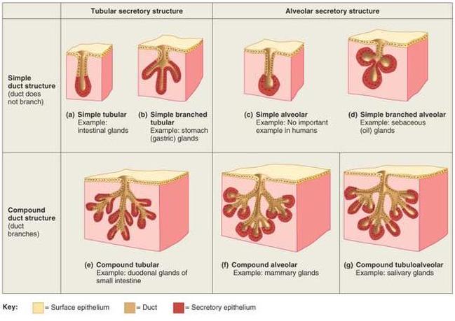
Multicellular Exocrine Glands- Each multicellular exocrine gland has two basic parts: an epithelium-walled duct and a secretory unit consisting of the secretory epithelium. Multicellular glands are classified by the structure of their ducts. Simple glands have unbranched ducts, whereas compound glands have a branched duct. The glands are further categorized by their secretory units: they are tubular if their secretory cells form tubes and alveolar if the secretory cells form spherical sacs. Furthermore, some glands are tubuloalveolar; that is, they contain both tubular and alveolar units.
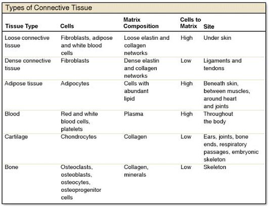
Connective Tissue- The most diverse and abundant type of tissue. There are four main classes of connective tissue and many subclasses. The main classes are: 1.) connective tissue proper 2.) cartilage 3.)bone tissue 4.) blood. Connective tissue do far more than just connect the tissues and organs of the body together. They also form the basis of the skeleton (bone and cartilage), store and carry nutrients (fat tissue and blood), surround all the blood vessels and nerves of the body (connective tissue proper), and lead the body's fight against infection.
Special Characteristics of Connective Tissue: relatively few cells, lots of extracellular matrix, extracellular matrix is composed of ground substance and fibers, embryonic origin.
Cells of the Connective Tissue: In most connective tissues, the primary cell type produces the extracellular matrix. In connective tissue proper, these cells are called fibroblasts. Fibroblasts make the protein subunits of fiber, which then assemble into fibers when the fibroblast secretes them. Fibroblasts also secrete the molecules that form the ground substance of the matrix. In cartilage tissue, the cells that secrete the matrix are called chondroblasts, and in bones they are osteoblasts. Once these tissue-forming cells are not actively secreting new matrix, they are termed fibrocytes, chondrocytes, and osteocytes. They function to maintain the tissue matrix and keep the tissue healthy.
Fibers of the Connective Tissue: The extracellular matrix of connective tissue is composed of fibers and ground substance. The three types of fibers found in connective tissue are: collagen fibers, reticular fibers, and elastic fibers. Collagen Fibers resist tension (pulling forces) and contribute strength to connective tissue. Reticular Fibers- are bundles of a special type of collagen fibril. These short fibers cluster into a mesh like network that covers and supports the structures bordering the connective tissue. Elastic Fibers- contain the rubberlike protein elastin, which allows them to function like rubber bands. Elastic fibers recoil.
Ground Substance: The molecules that compose the ground substance are produced and secreted by the primary cell type of the connective tissue (fibroblasts, chondroblasts, or osteoblasts). The ground substance in most connective tissues is a gel-like material that consists of large sugar and sugar-protein molecules (proteoglycans and glycosaminoglycans). The fluid-filled ground substance functions to cushion and protect body structures, to withstand compressive stresses, or to hold the tissue fluid that bathes all the cells in our body.
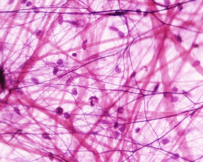 Areolar Loose Connective TIssue Proper
 Areolar Loose Connective Tissue Proper
Function: Loose packing, support, and nourishment for the structures with which it is associated
Location: Widely distributed throughout the body; substance on which epithelial basement membranes rest; packing between glands, muscles, and nerves; attaches the skin to underlying tissues.
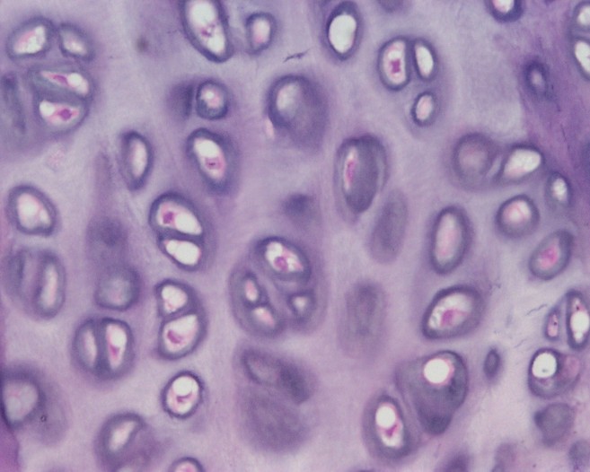 Elastic Cartilage
 Elastic Cartilage
Function: Provides rigidity with even more flexibility than hyaline cartilage because elastic fibers return to their original shape after being stretched.
Location: External ears, epiglottis, auditory tubes
Muscle Tissues
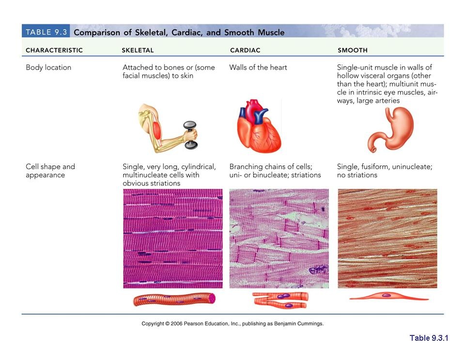
Muscle Tissue- The main characteristic of muscle tissue is its ability to contract or shorten, making movement possible. Muscle contraction results from contractile proteins located within the muscle cells. Muscle cells are also called muscle fibers. The three types of muscle tissue are: skeletal, cardiac, and smooth.
Skeletal muscle is what we normally think of as "muscle". Skeletal muscle attaches to the skeleton and enables the body to move. Skeletal muscle is described as voluntary because a person can purposefully cause skeletal muscle contraction to achieve specific body movements.
Cardiac muscle is the muscle of the heart. It is responsible for pumping blood. Cardiac muscle is under involuntary control, although a person can learn to influence the heart rate by using techniques such as meditation and biofeedback. Cardiac muscles are cylindrical but much shorter than skeletal muscle cells. Cardiac muscles are striated and usually have one nucleus per cell. They are often branched and connected to one another by intercalated discs. The intercalated discs, which contain specialized gap junctions, are important in coordinating the contractions of cardiac muscle cells.
Smooth muscle forms the walls of hollow organs (except the heart); it is also found in the skin and the eyes. Smooth muscle is responsible for a number of functions, such as moving food through the digestive tract and emptying the urinary bladder. Like cardiac muscle, smooth muscle is controlled involuntarily. Smooth muscle cells are tapered at each end, having a single nucleus, and are not striated.
Nervous Tissue
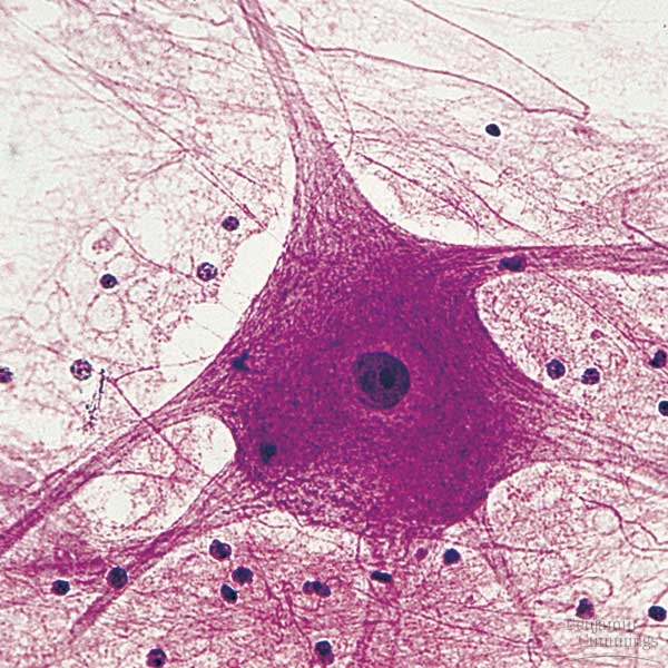
Nervous Tissue- forms the brain, spinal cord, and nerves. It is responsible for coordinating and controlling many body activities. Nervous tissue consists of neurons and support cells. The neuron, or nerve cell, is responsible for conducting action potentials. It is composed of three parts: a cell body, dendrites, and an axon. The cell body contains the nucleus and is the site of general cell functions. Dendrites and axons are nerve cell processes. Dendrites usually receive stimuli leading to electrical changes that either increase or decrease action potentials in the neuron's axon. Action potentials usually originate at the base of an axon where it joins the cell body and travel to the end of the axon. Neuroglia are the support cells of the nervous system; they nourish, protect, and insulate the neurons.
Function: Neurons transmit information in the form of action potentials, store information and integrate and evaluate data; neuroglia support, protect, and form specialized sheaths around axons.
Location: In the brain, spinal cord, and ganglia
Function: Neurons transmit information in the form of action potentials, store information and integrate and evaluate data; neuroglia support, protect, and form specialized sheaths around axons.
Location: In the brain, spinal cord, and ganglia
 RSS Feed