
Sense is the ability to perceive stimuli. The senses are the means by which the brain receives information about the environment and the body. Sensation, or perception, is the conscious awareness of stimuli received by sensory receptors.
Historically five senses are recognized: smell, taste, sight, hearing, and touch. Today we recognize many more senses and divide them into two basic groups:general and special senses. The general senses have receptors distributed over a large part of the body. They are divided into two groups: the somatic senses and the visceral senses. The somatic senses provide sensory information about the body and the environment. The visceral senses provide information about various internal organs, primarily involving pain and pressure.
Special senses are more specialized in the structure and are localized to specific parts of the body. THe special senses are sell, taste, sight, hearing, and balance.
General Senses
The General senses are widely distributed though out the body an include the senses of touch, pressure, pain, temperature, vibration, itch, and proprioception,, which is the sense of movement and position of the body and limbs.
Sensory receptors are sensory nerve endings or specialized cells capable of responding to stimuli by developing action potentials. Several types of receptors are associated with both the social and the general senses, and each responds to a different type of stimulus:
Mechanoreceptors respond to mechanical stimuli, such as the bending or stretching of receptors.
Chemoreceptors respond to chemicals, such as odor molecules.
Photoreceptors respond to light.
Thermoreceptors respond to temperature changes.
Nociceptors respond to stimuli that result in the sensation of pain.
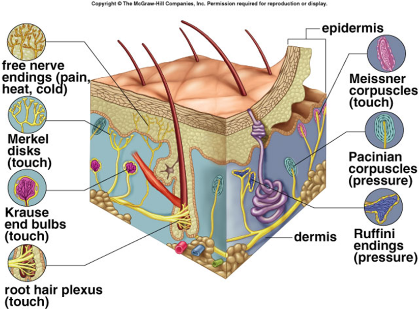
Free Nerve Endings are the most common receptors and are relatively unspecialized neuronal branches that are similar to dendrites. Free nerve endings are distributed throughout almost all parts of the body. Some free nerve endings respond to painful stimuli, some to temperature, some to itch, and some to movement.
Cold Receptors or Warm Receptors are receptors for temperature. Cold receptors respond to decreasing temperatures but stop responding at temperatures below 12 C. Warm receptors respond to increasing temperatures but stop responding at temperatures about 47 C. It is sometimes difficult to distinguish very cold from very warm objects touching the skin because only pain receptors are stimulated at temperatures below 12 C or above 47 C.
Touch Receptors are structurally more complex than free nerve endings, and many are enclosed by capsules. There are several types of touch receptors: Merkel disks are small, superficial nerve endings involved in detecting light touch and superficial pressure. Hair follicle receptors, associated with hairs, are also involved in detecting light touch. LIght touch receptors are very sensitive but not very discriminative, meaning that the point being touched cannot be precisely located. Receptors for fine, discriminative touch, called Meissner corpuscles, are located just deep to the epidermis. These receptors are very specific in localizing tactile sensations. Deeper tactile receptors, called Ruffini corpuscles play an important role in detecting continuous pressure in the skin. The deepest receptors are associated with tendons and joints and are called pacinian corpuscles. These receptors relay information concerning deep pressure, vibration, and position (proprioception).
Special Senses
The senses of smell, taste, sight, hearing, and balance are associated with very specialized, localized sensory receptors. The sensations of smell and taste are closely related, both structurally and functionally, and both are initiated by the interaction of chemicals with sensory receptors. The sense of vision is initiated by the interaction of light with sensory receptors. Both hearing and balance function in response to the interaction of mechanical stimuli with sensory receptors. Hearing occurs in response to sound waves, and balance occurs in response to gravity or motion.
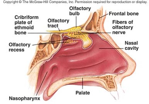
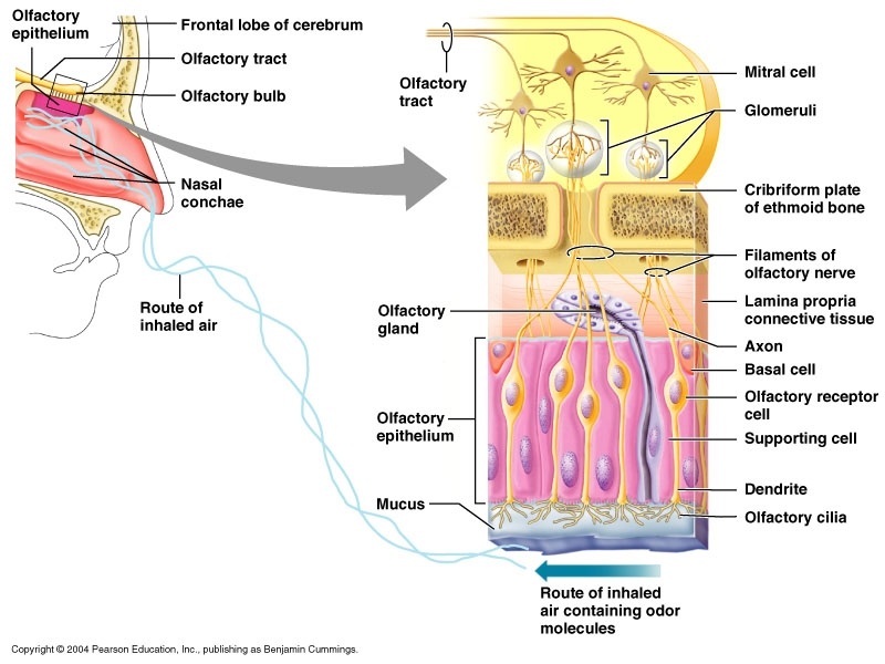
Olfaction is the sense of smell, and occurs in response to airborne molecules, called odorants, that enter the nasal cavity. Olfactory neurons are bipolar neurons within the olfactory epithelium, which lines the superior part of the nasal cavity.
Axons from olfactory neurons form the olfactory nerves (cranial nerve I), which pass through foramina of the cribiform plate and enter the olfactory bulb. There they synapse with interneurons that relay action potentials to the brain through the olfactory tracts. Each olfactory tract terminates in an area of the brain called the olfactory cortex, located within the temporal and frontal lobes. Olfaction is a major sensation that is relayed directly to the cerebral cortex without first passing through the thalamus.
The senses of smell, taste, sight, hearing, and balance are associated with very specialized, localized sensory receptors. The sensations of smell and taste are closely related, both structurally and functionally, and both are initiated by the interaction of chemicals with sensory receptors. The sense of vision is initiated by the interaction of light with sensory receptors. Both hearing and balance function in response to the interaction of mechanical stimuli with sensory receptors. Hearing occurs in response to sound waves, and balance occurs in response to gravity or motion.
Olfaction is the sense of smell, and occurs in response to airborne molecules, called odorants, that enter the nasal cavity. Olfactory neurons are bipolar neurons within the olfactory epithelium, which lines the superior part of the nasal cavity.
Axons from olfactory neurons form the olfactory nerves (cranial nerve I), which pass through foramina of the cribiform plate and enter the olfactory bulb. There they synapse with interneurons that relay action potentials to the brain through the olfactory tracts. Each olfactory tract terminates in an area of the brain called the olfactory cortex, located within the temporal and frontal lobes. Olfaction is a major sensation that is relayed directly to the cerebral cortex without first passing through the thalamus.
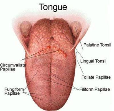
The sensory structures that detect taste stimuli are the taste buds. Taste buds are oval structures located on the surface of certain papillae, which are enlargements on the surface of tongue. Taste buds are also distributed throughout other areas of the mouth and pharynx, such as on the palate, the root of the tongue, and the epiglottis. Each taste bud consists of two types of cells. Specialized epithelial cells form the exterior supporting capsule of each taste bud, and the interior consists of about 40 taste cells. Each taste cell contains hairlike processes, called taste hairs, that extend into a tiny opening in the surrounding stratified epithelium, called a taste pore.
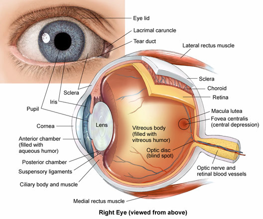
Eyelids with their associated lashes, protect the eyes from foreign objects. If an object suddenly approaches the eye, the eyelids protect the eye by closing and then opening quite rapidly (blink reflex). Blinking, which normally occurs about 20 times per minute, also helps keep the eyes lubricated by spreading tears over the surface.
The conjunctiva is a thin, transparent mucous membrane covering the inner surface of the eyelids and the anterior surface of the eye. The secretions of the conjunctiva help lubricate the surface of the eye. Conjunctivitis is an inflammation of the conjunctiva.
The lacrimal apparatus consists of a lacrimal gland situated in the superior lateral corner of the orbit and a nasolacrimal duct and associated structures in the inferior medial corner of the orbit. The lacrimal gland produces tears, which pass over the anterior surface of the eye. Most of the fluid produced by the lacrimal glands evaporates from the surface of the eye, but excess tears are collected in the medial angle of the eyes by small ducts called lacrimal canaliculi. These canaliculi open into a lacrimal sac, an enlargement of the nasolacrimal duct, which opens into the nasal cavity. Tears lubricate and cleanse the eye. They also contain an enzyme that helps combat eye infections.
Movement of each eyeball is accomplished by six skeletal muscles called the extrinsic eye muscle. For of these muscles run more or less straight from their origins in the posterior portion of the orbit to their insertion sites on the eye, to attach to the four quadrants of the eyeball. They are the superior, inferior, medial, and lateral rectus muscles. Two muscles, the superior and inferior oblique muscles, are located to an angle to the long axis of the eyeball.
The outer segments of rod and cone cells are modified by numerous foldings of the cell membrane to form discs. Rod cells contain photosensitive pigment called rhodopsin, which is made up of the colorless protein opsin in loose chemical combination with a yellow pigment called retinal.
Visual Pathways
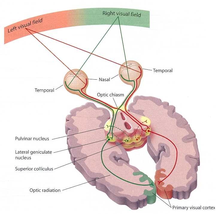
Neuronal Pathways for Vision
- Each visual field is divided into temporal and nasal halves.
- After passing through the lens, light from each half of a visual fields projects to the opposite side of the retina.
- An optic nerve consists of axons extending from the retina to the optic chasm.
- In the optic chasm, axons from the nasal part of the retina cross and project to the opposite side of the brain. Axons from the temporal part of the retina do not exist.
- An optic tract consists of axons that have passed through the optic chasm (with or without crossing) to the thalamus.
- The axons synapse in the thalamus. Collateral branches of the axons in the optic tracts synapse in the superior colliculi.
- An optic radiation consists of axons from thalamic neurons that project to the visual cortex.
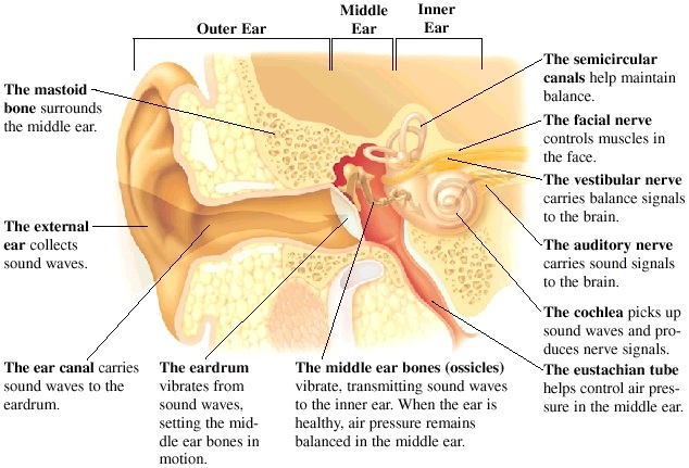
Effect of Sound Waves o Middle and Inner Ear Structures
Hearing
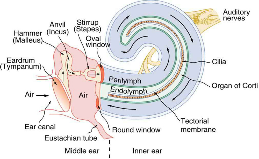
- Sound waves strike the tympanic membrane and cause it to vibrate
- Vibration of the tympanic membrane causes the malleus, the incus, and the stapes to vibrate
- The foot plate of the stapes vibrates in the oval window
- Vibration of the foot palate causes the perilymph in the scala vestibule to vibrate
- Vibration of the perilymph causes the vestibular membrane to vibrate, which causes vibrations in the endolymph.
- Vibration of the endolymph causes displacement of the basilar membrane. Short waves (high pitch), causes displacement of the basilar membrane near the coal window, and longer waves (low pitch) cause displacement of the basilar membrane some distance from the oval window. Movement of the basilar membrane is detected in the hair cells of the spiral organ, which are attached to the basilar membrane. Vibrations of the perilymph in the scala vestibuli and of the basilar membrane are transferred to the perilymph of the scala tympani.
- Vibrations in the perilymph of the scala tympani are transferred to the round window, where they are dampended.
 RSS Feed