Functions of the Respiratory System:
- Regulation of blood pH
- Voice production
- Olfaction
- Innate immunity
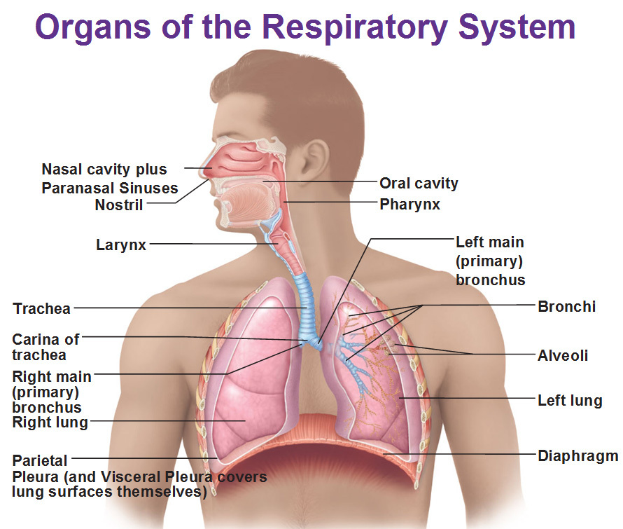
The Respiratory System is divided into the upper and lower respiratory tract. The upper respiratory tract includes: the external nose, the nasal cavity, the pharynx, and associated structures. The lower respiratory tract includes: the larynx, the trachea, the bronchi, and the lungs.
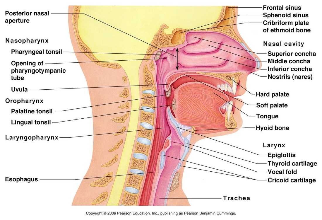
The pharynx is the common passageway for both the respiratory and the digestive system. Air from the nasal cavity and air, food, and water from the mouth pass through the pharynx. Inferiorly, the pharynx leads to the rest of the rest of the respiratory system through the opening into the larynx and to the digestive system through the esophagus. The pharynx can be divided into three regions: the nasopharynx, the oropharynx, and the laryngopharynx.
The larynx is located in the anterior throat and extends from the base of the tongue to the trachea. It is a passageway for air between the pharynx and the trachea. The larynx consists of an outer casing of nine cartilages connected to one another by muscle and ligaments.
The trachea, or windpipe, is a membranous tube attached to the larynx. It consists of connective tissue and smooth muscle, reinforced with 16-20 C-shaped pieces of hyaline cartilage.
The trachea divides into the left and right main bronchi, or primary bronchi, each of which connects to a lung. The left main bronchus is more horizontal than the right main bronchus because it is displaced by the heart. Foreign objects that enter the trachea usually lodge in the right main bronchus, because it is wider, shorter, and more vertical than the left main bronchus and is more in direct line with the trachea. The main bronchi extend from the trachea to the lungs.
The lungs are the principal organs of respiration. Each lung is cone-shaped, with its base resting on the diaphragm and its apex extending superiorly to a point about 2.5 cm above the clavicle. The right lung has three lobes, called the superior, middle and inferior lobes. The left lung has two lobes, called the superior and inferior lobes. The lobes of the lungs are separated by deep, prominent fissures on the lung surface.
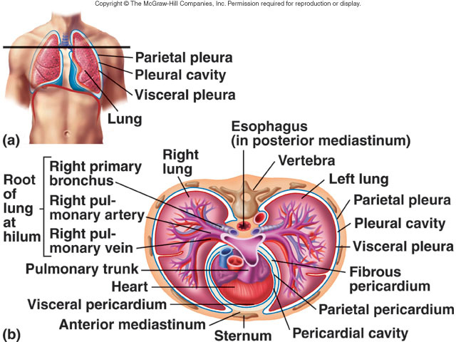
Pleural Cavities:
The lungs are contained within the thoracic cavity. In addition, each lung is surrounded by a separate pleural cavity. Each pleural cavity is lined with a serous membrane called the pleura. The pleura consists of a parietal and a visceral part. The parietal pleura, which lines the walls of the thorax, diaphragm, and mediastinum, is continuous with the visceral pleura, which covers the surface of the lung.
The lungs are contained within the thoracic cavity. In addition, each lung is surrounded by a separate pleural cavity. Each pleural cavity is lined with a serous membrane called the pleura. The pleura consists of a parietal and a visceral part. The parietal pleura, which lines the walls of the thorax, diaphragm, and mediastinum, is continuous with the visceral pleura, which covers the surface of the lung.
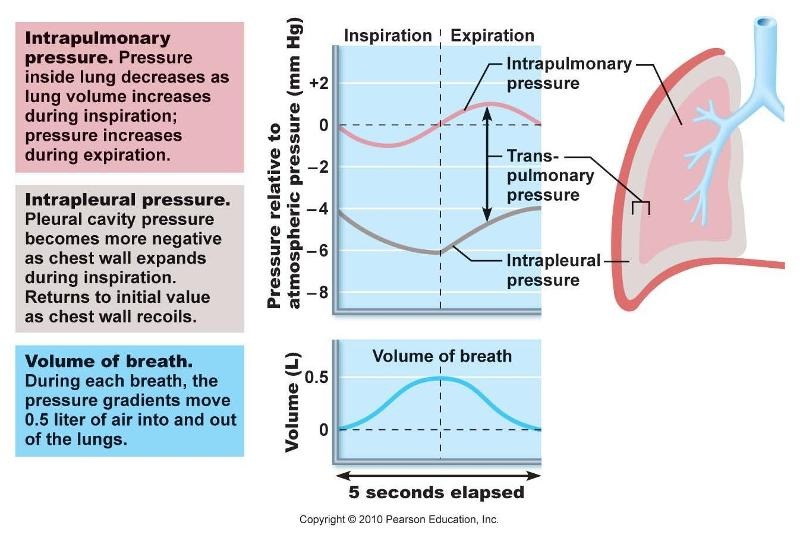
Respiratory Volumes:
- Tidal Volume- the volume of air inspired or expired with each breath. At rest, quiet breathing results in a tidal volume of about 500 mL.
- Inspiratory reserve volume- the amount of air that can be inspired forcefully beyond the resting tidal volume. (about 3000 mL)
- Expiratory reserve volume- the amount of air that can be expired forcefully beyond the resting tidal volume (about 1100 mL)
- Residual volume- the volume of air still remaining in the respiratory passages and lungs after maximum expiration (about 1200 mL)
Respiratory Capacities:
- Functional residual capacity- the expiratory reserve volume plus the residual volume. This is the amount of air remaining in the lungs at the end of a normal expiration (about 2300 mL at rest)
- Inspiratory capacity- the tidal volume plus the inspiratory reserve volume. This is the amount of air a person can inspire maximally after a normal expiration (about 3500 mL)
- Vital capacity- the sum of the inspiratory reserve volume, the tidal volume, and the expiratory reserve volume. It is the maximum volume of air that a person can expel from the respiratory tract after a maximum inspiration (about 4600 mL)
- Total lung capacity- the sum of the inspiratory and expiratory reserves and the tidal and residual volumes (about 5800 mL). The total lung capacity is also equal to the vital capacity plus the residual volume.
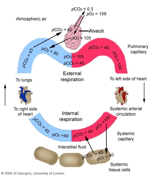
Gas Exchange:
- Oxygen diffuses into the arterial ends of pulmonary capillaries, and CO2 diffuses into the alveoli because of differences in partial pressures.
- As a result of diffusion at the venous ends of pulmonary capillaries, the Po2 in the blood is equal to the Po2 in the alveoli, and the Pco2 in the blood is equal to the Pco2 in the alveoli.
- The Po2 of blood in the pulmonary veins is less than in the pulmonary capillaries because of mixing with deoxygenated blood from veins draining the bronchi and bronchioles.
- Oxygen diffuses out of the arterial ends of tissue capillaries, and CO2 diffuses out of the tissue because of differences in partial pressures.
- As a result of diffusion at the venous ends of tissue capillaries, the Po2 in the blood is equal to the Po2 in the tissue, and the Pco2 in the blood is equal to the Pco2 in the tissue.
 RSS Feed