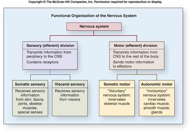
Functions of the Nervous System
- Receiving sensory input. Sensory receptors monitor numerous external and internal stimuli.
- Integrating information. The brain and spinal cord are the major organs for processing sensory input and initiating responses. The input may produce an immediate response, be stored as memory, or be ignored.
- Controlling muscles and glands. Skeletal muscles normally contract only when stimulated by the nervous system. Thus, by controlling skeletal muscle, the nervous system controls the major body movements of the body. The nervous system also participates in controlling cardiac muscle, smooth muscle, and many glands.
- Maintaining homeostasis. This function depends on the nervous system's ability to detect, interpret, and respond to changes in internal and external conditions. In response, the nervous system can stimulate or inhibit the activities of other systems to help maintain a constant internal environment.
- Establishing and maintaining mental activity. The brain is the center of mental activity, including consciousness, memory, and thinking.
Cells of the Nervous System
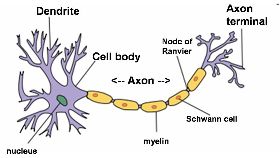
Neurons- or nerve cells, receive stimuli, conduct action potentials and transmit signals to other neurons or effector organs. There are three parts to a neuron: a cell body and two types of processes, called dendrites and axons.
Each neuron cell body contains a single nucleus. As with many other cell, the nucleus of the neuron is the source of information for gene expression.
Dendrites- are short, often highly branching cytoplasmic extensions that are tapered from their bases at the neuron cell body to their tips. Most dendrites are extensions of the neuron cell body, but dendritelike structures also project from the peripheral ends of some sensory axons. Dendrites usually receive information from other neurons or from sensory receptors and transmit the information toward the neuron cell body.
Each neuron has an axon, a single long cell process extending from the neuron cell body. The area where the axon leaves the neuron cell body is called the axon hillock. Each axon has a uniform diameter and may vary in length from a few millimeters to more than a meter. Axons of sensory neurons conduct action potentials towards towards the CNS, and axons of motor neurons conduct action potentials from one part of the brain or spinal cord to another part. An axon may remain unbranched or may branch to form collateral axons. Axons can be surrounded by highly specialized insulating later of cells called the myelin sheath.
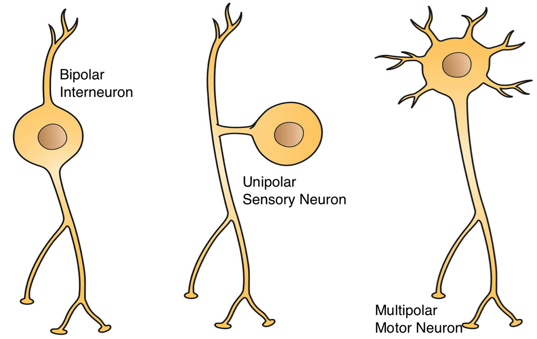
Types of Neurons:
Multipolar neurons- have many dendrites and a single axon.
Bipolar neurons- have two processes: one dendrite and one axon. Bipolar neurons are located in some sensory organs, such as in the retina of the eye and in the nasal cavity.
Pseudo-unipolar neurons- have a single process extending from the cell body. This process divides into two processes a short distance from the cell body. One process extends to the periphery, and the other extends to the CNS. The two extensions function as a single axon with small, dendrite like sensory receptors at the periphery. The axon receives sensory information at the periphery and transmits that information in the form of action potentials to the CNS.
The Resting Membrane Potential is generated by three main factors: 1.) a higher concentration of K+ immediately inside the cell membrane, 2.) a higher concentration of Na+ immediately outside the cell membrane, and 3.) greater permeability of the cell membrane to K+ than to Na+. Thus, the resting membrane potential results from differences in the concentration of ions across the membrane and the permeability characteristics of the membrane.
A Synapse is a junction where the axon of one neuron interacts with another neuron or with cells of an effector organ, such as a muscle or gland. The end of the axons forms a presynaptic terminal. The membrane of the dendrite or effector cell is the postsynaptic membrane, and the space separating the presynaptic and postsynaptic membranes is the synaptic cleft. Chemical substances called neurotransmitters are stored in synaptic vesicles in the presynaptic terminal.
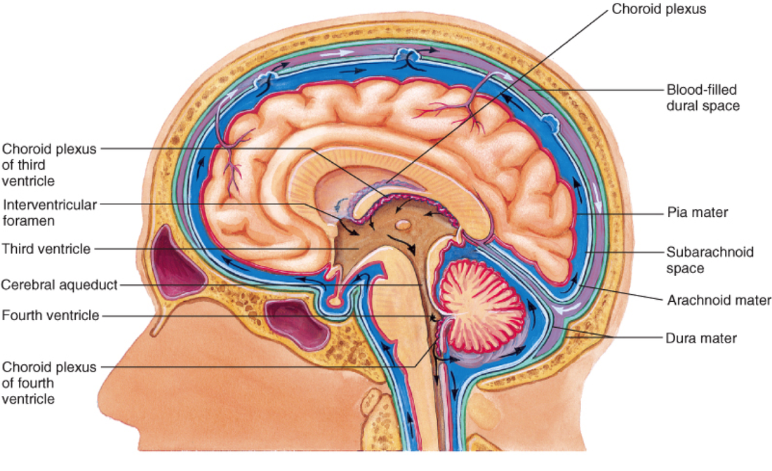
Flow of Cerebrospinal Fluid
- Cerebrospinal Fluid (CSF) is produced by the choroid plexuses of each of the four ventricles (inset, lower left).
- CSF from the lateral ventricles flows to the third ventricle.
- CSF flows from the third ventricle through the cerebral aqueduct to the fourth ventricle.
- CSF exits the fourth ventricle through openings in the wall of the fourth ventricle and enters the subarachnoid space. Some CSF enters the central canal of the spinal fluid.
- CSF flows through the subarachnoid space to the arachnoid granulations in the superior sagittal sinus, where it enters the venous circulation (inset, upper right).
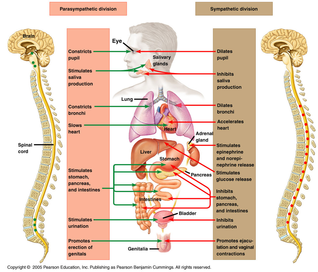
The autonomic nervous system is composed of the sympathetic and the parasympathetic division. Increased activity in sympathetic neurons generally prepares the individual for physical activity, whereas parasympathetic stimulation generally activates involuntary functions, such as digestion, that are normally associated with the body at rest.
 RSS Feed