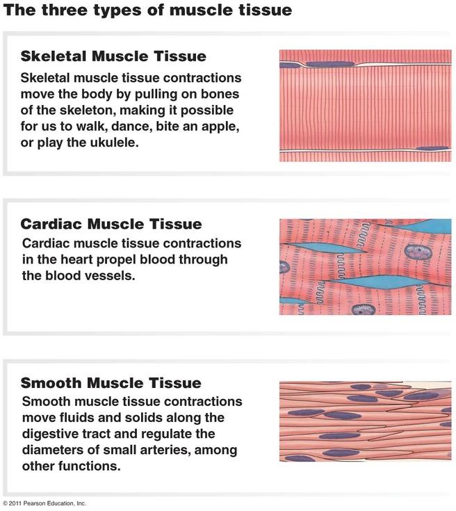
Functions of the Muscular System
- Movement of the body
- Maintenance of posture
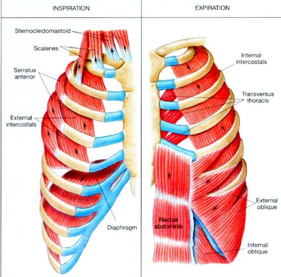
- Respiration
- Production of body heat
- Communication
- Constriction of organs and vessels
- Contraction of the heart
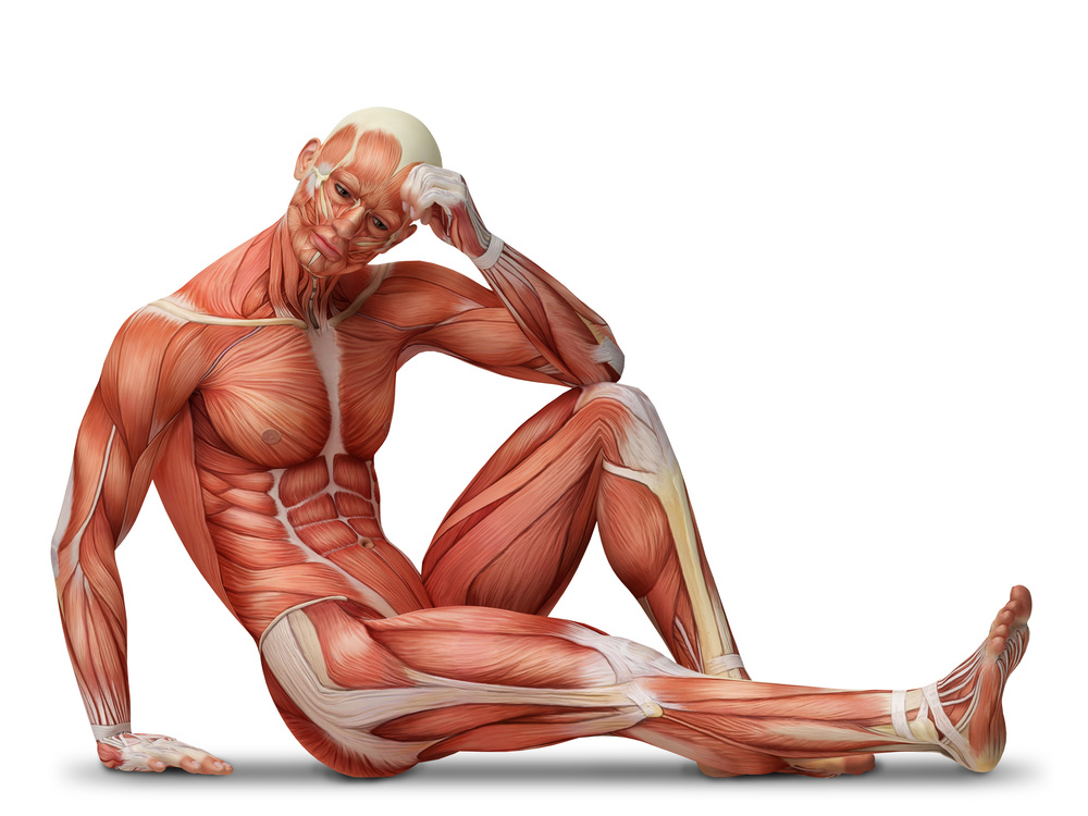
Characteristics of Skeletal Muscle
- Contractibility. When skeletal muscles contract, they cause the structures to which they are attacked to move. Skeletal muscles shorten forcefully during contraction, but they lengthen passively. EIther gravity or the contraction of an opposing muscle produces a force that pulls on the shortened muscle, causing it to lengthen.
- Excitability. The capacity of skeletal muscle to respond to a stimulus. Normally, the stimulus is from nerves that we consciously control.
- Extensibility. The skeletal muscles stretch. After a contraction, skeletal muscles can be stretched to their normal resting length and beyond to a limited degree.
- Elasticity. Skeletal muscles are able to recoil to their original resting length after they have been stretched.
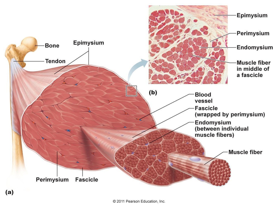
Connective TIssue Coverings of Muscle
Each skeletal muscle is surrounded by a connective tissue sheath called the epimysium or muscular fascia. A muscle is composed of numerous visible bundles called muscle fasciculi, which are surrounded by loose connective tissue called the perimysium. A fascicles is composed of several muscle cells, which are called muscle fibers. Each muscle fiber is surrounded by loose connective tissue called endomysium.
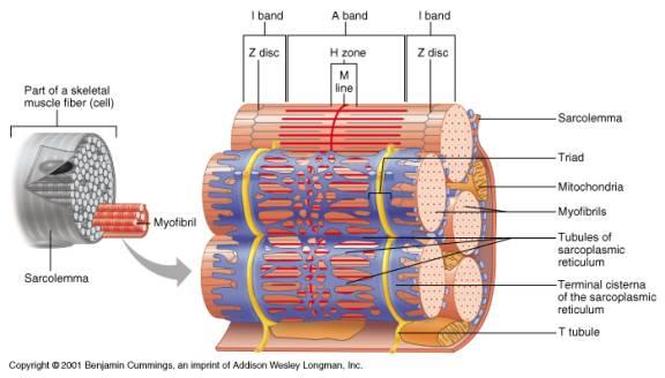
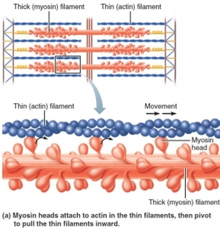
Under the light microscope, the light and dark striations in skeletal muscle fibers are clearly visible. These striations result from the internal structure of long, rod-shaped organelles called myofibrils. Myofibrils are unbranched cylinders that are present in large numbers, making up more than 80% of the sarcoplasm. They are specialized in contractile organelles unique to muscle tissue. The myofibrils in a fiber are separated from one another by other components of a sarcoplasm. Among those components are mitochondria and glycosides, both of which supply energy for muscle contraction. A myofibril is a long row of repeating segments called sacromeres. The sacromere is the basic unit of contraction skeletal muscle. The boundaries at the two ends of each sacromere are called Z discs. Attached to each Z disc and extending toward the center of the sacromere are many fine myofilaments called thin (actin) filaments, which consist primarily of the protein actin, although they contain other proteins as well. In the center of the sacromere and overlapping the inner ends of the thin filaments is a cylindrical bundle of thick (myosin) filaments. Thick filaments consist largely of myosin molecules. THey also contain ATPase enzymes that split ATP to release the energy required for muscle contraction. Both ends of thick filaments are studded with knobs called myosin heads.
The Sacromere structure explains the pattern of striations in skeletal muscle fibers. The dark bands are created by the full length of the thick filaments in the sacromeres, along with the inner ends of the thin filaments, which overlap the thick filaments. This region of each sacromere, is called the A band. The central part of an A band, where no thin filaments reach, is the H Zone. The M line in the center of the H zone contains tiny rods that hold the thick filaments together. THe two regions on either side of the A band, regions that contain only thin filaments, are called the I bands. It is the I bands of the sacromeres that create the light portions of the light-dark pattern of striations seen along the length of any skeletal muscle fiber.
The Sacromere structure explains the pattern of striations in skeletal muscle fibers. The dark bands are created by the full length of the thick filaments in the sacromeres, along with the inner ends of the thin filaments, which overlap the thick filaments. This region of each sacromere, is called the A band. The central part of an A band, where no thin filaments reach, is the H Zone. The M line in the center of the H zone contains tiny rods that hold the thick filaments together. THe two regions on either side of the A band, regions that contain only thin filaments, are called the I bands. It is the I bands of the sacromeres that create the light portions of the light-dark pattern of striations seen along the length of any skeletal muscle fiber.
There are two types of muscle contraction involved in producing movement: concentric contraction and eccentric contraction. Concentric contraction is the more familiar type, in which the muscle shortens and does work- picking up a book or kicking a ball. Eccentric contraction- occurs when a muscle generates force as it lengthens. The mechanism for this type of contraction is less understood, but eccentric contractions are essential for controlled movement and resistance to gravity.
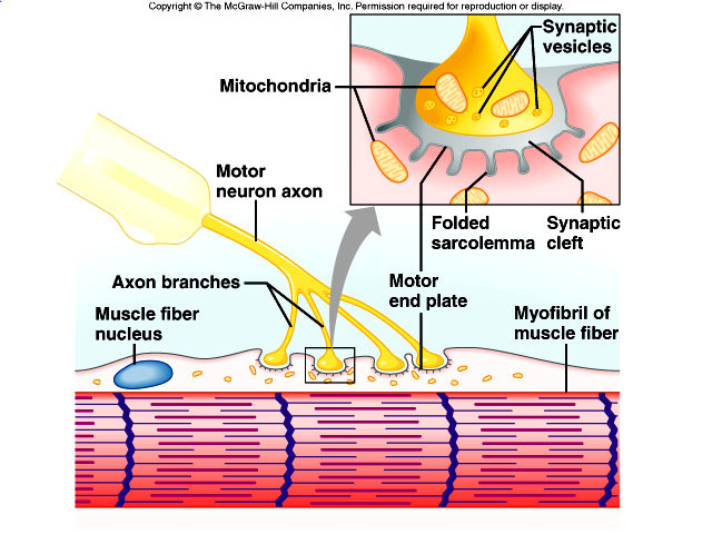
The release of calcium ions from the sarcoplasmic reticulum and the subsequent contraction of skeletal muscle is initiated by nervous stimulation. THe nerve cells that innervate muscle fibers are called motor neurons. Each muscle fiber in a skeletal muscle is served by a nerve ending, which signals the fiber to contract. The point at which the nerve ending and fiber meet is called a neuromuscular junction. The nerve part of the junction is a cluster of enlargements at the end of the axonal process that stores chemical messenger molecules, neurotransmitters. These enlargements are called axon terminals. The axon terminals are separated from the sarcolemma of the muscle fiber by a space called the synaptic cleft. THe axon terminals contain vesicles that release neurotransmitters when a nerve impulse reaches the terminals. The neurotransmitters at the neuromuscular junction-acetylcholine-diffuses across the synaptic cleft and binds to receptor molecules on the sarcolemma, where it induces an impulse that initiates fiber contraction.
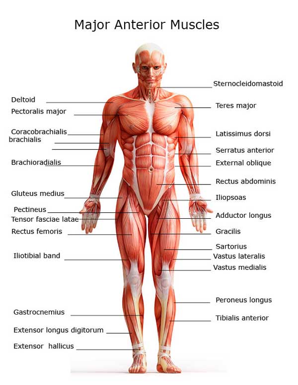
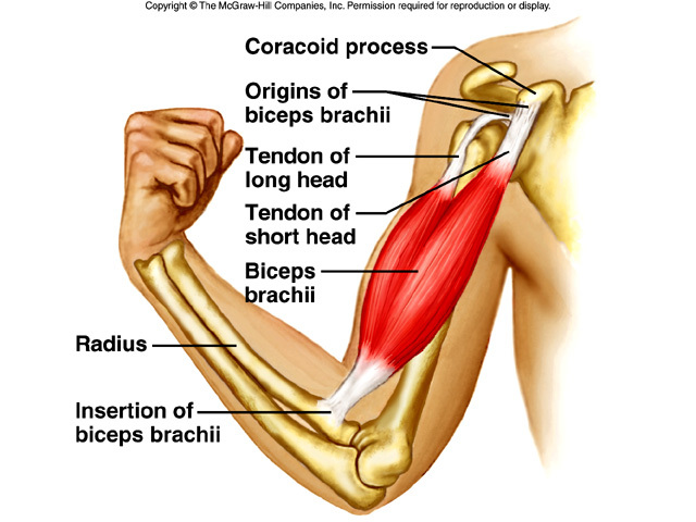
The two points of attachment of each muscle are its origin and insertion. The origin, also called the head, is the most stationary end of the muscle. The insertion is the end of the muscle attached to the bone undergoing the greatest movement. Origins are usually, but not always, proximal or medial to the insertion of a given muscle. THe part of the muscle between the origin and the insertion is the belly.
Muscles are typically grouped so that the action of one muscle or group of muscles is opposed by that of another muscle or group of muscles. A muscle that accomplishes a certain movement, such as flexion, is called the agonist. A muscle acting in opposition to an agonist is called an antagonist. Members of a group of muscles working together to produce a movement are called synergists. Among a group of synergists, if one muscle plays the major role in accomplishing the desired movement, it is called the prime mover. Fixators are muscles that hold one bone in place relative to the body while a usually more distal bone is moved.
Muscles are typically grouped so that the action of one muscle or group of muscles is opposed by that of another muscle or group of muscles. A muscle that accomplishes a certain movement, such as flexion, is called the agonist. A muscle acting in opposition to an agonist is called an antagonist. Members of a group of muscles working together to produce a movement are called synergists. Among a group of synergists, if one muscle plays the major role in accomplishing the desired movement, it is called the prime mover. Fixators are muscles that hold one bone in place relative to the body while a usually more distal bone is moved.
 RSS Feed