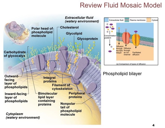
The membrane is composed of a bilayer of phospholipids and cholesterol with proteins "floating" in the membrane. The nonpolar hydrophobic region of each phospholipid molecule is directed toward the center of the membrane, and the polar hydrophilic region is directed toward the fluid environment either outside or inside the cell.
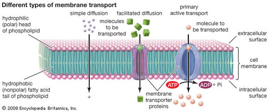
Simple Diffusion- Small, uncharged molecules, such as oxygen, carbon dioxide, and fat-soluble molecules, can pass freely through the lipid bilayer of the plasma membrane. Osmosis- The diffusion of water molecules across a membrane. Facilitated Diffusion- Water-soluble or charged molecules, such as glucose, amino acids, and ions, cannot pass through the lipid bilayer by simple diffusion. Such substances can cross the plasma membrane only by means of specific transport mechanisms that use integral proteins to carry or pump molecules across the membrane or to form channels through which specific molecules pass. Active Transport- The movement of molecules across the plasma membrane across their concentration gradient.
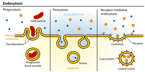
Phagocytosis- "Cell eating." The cell engulfs a large particle by forming pseudopods ("false feet") around it and enclosing it within a membrane sac called a phagosome. THe phagosome then combines with a lysosome, and its contents are digested. Vesicle may or may not be protein-coated but has receptors capable of binding to microorganisms or solid particles. Pinocytosis- The cell "gulps" drops of extracellular fluid containing solutes into tiny vesicles. No receptors are used, so the process is nonspecific. Most vesicles are protein-coated. Receptor-Mediated Endocytosis- Extracellular substances bind to specific receptor protein-coated pits, enabling the cell to ingest and concentrate specific substances in protein-coated vesicles. The ingested substance may simply be released inside the cell, or combined with a lysosome to digest contents. Receptors are recycled to the plasma membrane in vesicles.
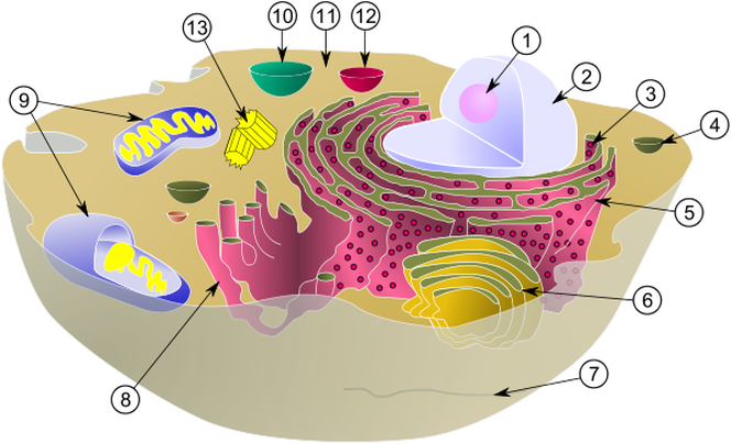
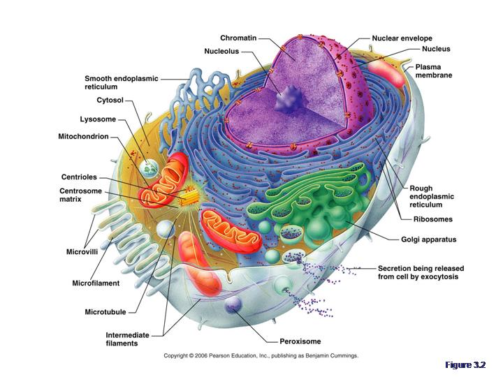
Match the Organelle with its function!
A. Site of Protein Synthesis
B. Makes proteins that are secreted from the cell; makes the cell's membranes
C. Site of lipid synthesis, lipid metabolism, and drug detoxification
D. Packages, modifies, and segregates proteins for secretion from the cell, inclusion in lysosomes, and incorporation into the plasma membrane
E. Sites of intracellular indigestion
F. Sites of intracellular digestion
G. Site of ATP synthesis; powerhouse of the cell
H. The enzymes detoxify a number of toxic substances; the most important enzyme, catalase, breaks down hydrogen peroxide
I. Involved in muscle contraction and other types of intracellular movement; help form the cell's cytoskeleton
J. Involved in muscle contraction and other types of intracellular movement; helps form the cell's cytoskeleton
K. The stable cytoskeleton elements; resist tension forces acting on the cell
L. Support the cell and give it shape; involved in intracellular and cellular movements; form centrioles
M. Organize a microtubule network during mitosis to form the spindle and asters; form the bases of cilia and flagella
N. Control center of the cell; responsible for transmitting genetic information and providing the instructions for protein synthesis
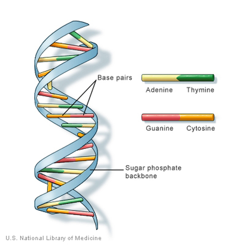
The DNA is a long double helix that resembles a spiral staircase. THis double helix is in turn composed of four kinds of subunits called nucleotides, each of which contains a distinct base. These bases- thymine, adenine, cytosine, and guanine- bind to form the "stairs" of the "staircase" and to hold the DNA helix together. The double helix of DNA is packed with protein molecules and coiled ins trends of increasing structural complexity and thickness. The DNA molecule plus the proteins form chromatin. Each two turns of the DNA helix is packed with eight disc-shaped protein molecules called histones. Each cluster of DNA and histones is called a nucleosome. In an electron micrograph of chromatin, the nucleosomes have the appearance of beads on a string. Chromatin in this form is called extended chromatin. Further coiling of the nucleosomes forms a tight helical fiber. THese thick fibers of chromatin are called condensed chromatin.
 RSS Feed